Apoptosis
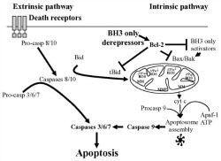
 Apoptosis is a genetic process by which a cell is programmed to die. This process is critical for development and proper function of an organism. Two main pathways lead to Apoptosis (right figure; Wlodkowic D, Met Cell Biol. 103, 2011): Extrinsic (activated mainly by death ligands, such TNFR, TRAIL, and FasL) and Intrinsic (activated by DNA damage, hypoxia, survival factor deprivation, etc.), resulting in the activation of a family of cysteine proteases, caspases. As a consequence of caspase activation a proteolytic cascade occurs leading to cell death and elimination of dying cells.
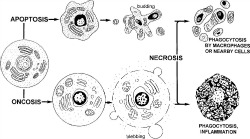
Morphological (cell volume fluctuations, phosphatidylserine “flipping”, loss of membrane permeability, chromatin condensation) and biochemical (mitochondrial potential changes, caspase, other signaling events) changes are characteristic of Apoptosis. Importantly, most of those changes can be detected and measured by flow cytometry (bottom fig; Darzynkiewicz Z, Cytometry 27, 1997).
However, depending on the cell t ype and its stage of differentiation/activation, the apoptotic process may have a lot of phenotypic variations. Then, to properly characterize Apoptosis always measure it using several different methods, preferably in the same sample. Multiparametric flow cytometry is ideal for this. Do not only measure cell death, but characterize it! (advice from Dr. Bill Telford, NCI Flow Cytometry Core Lab).