Asylum Atomic Force Microscope
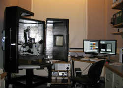
Atomic Force MicroscopeMFP-3D-BIOTM Atomic Force Microscope by Asylum Research  is mounted on Olympus IX71 inverted microscope that resides on a Herzan AVI 350-S Active Vibration Isolation Table which dynamically dampens the most extreme vibrational disturbances; and the system is totally enclosed in an air tight state-of-the-art BCH-45 Large Acoustic Isolation Hood with air temperature control capabilities. The closed looped system allows investigation of dry or hydrated biological/materials surface features at very high resolution to include dynamic 3D visual analysis, force curves, optical (fluorescence and phase) image overlays precisely registered over AFM topographical information; plus advanced modes such as nanomanipulation(s), nanoindentation(s), Dual AC and more. The system software modules are written in Igor Pro, an integrated program by Wavemetrics which provides very advanced features for visualizing, analyzing, transforming and presenting experimental data.


Cypher Atomic Force Microscope
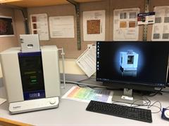
cypherAFM

The Asylum Research Cypher ES boasts exceptional performance and full environmental control features. It masters high resolution imaging with speed and stability while containing an easily operatable environmental chamber to control gas or liquid environments, at temperatures from 0-250°C, and in some of the harshest chemical environments. The Cypher ES is the ultimate AFM for the most demanding experimental requirements.

  • Routinely achieve higher resolution than other AFMs
  • Fast scanning with results in seconds instead of minutes
  • Every step of operation is simpler for remarkable productivity
  • Small footprint in the lab, huge potential to grow in capability
  • Support that goes above and beyond your expectations
  • Enables gas and liquid perfusion through a sealed cell
  • Controls sample temperature over a wide 0–250°C range
  • Broadest compatibility with harsh chemicals


Papers published from the MIC:

  • Braet, F., et al. (2018). "Probing the unseen structure and function of liver cells through atomic force microscopy." Semin Cell Dev Biol 73: 13-30.
  • Headrick, R. J., et al. (2018). "Structure-Property Relations in Carbon Nanotube Fibers by Downscaling Solution Processing." Adv Mater 30(9).
  • Headrick, R. L., et al. (2019). "Coherent X-ray measurement of step-flow propagation during growth on polycrystalline thin film surfaces." Nat Commun 10(1): 2638.
  • Li, Y., et al. (2016). "Nucleation and strain-stabilization during organic semiconductor thin film deposition." Sci Rep 6: 32620.
  • Li, Y., et al. (2016). "Measuring adhesion between modified asphalt bindrs and aggregate minerals: use of particle probe scanning force microscopes." Transportation Research Record.
  • Li, Y., et al. (2017). "Adhesion between modified binders and aggregate minerals at ambient conditions measured with particle-probe scanning force microscopes." Journal of Materials in Civil Engineering.
  • Li, Y., et al. (2017). "Statistical analyses of aggregate mineral-binder adhesion: measuring with particle probe scanning force microscopes." Transportation Research Record 2632(1): 25-21.
  • Li, Y., et al. (2018). "Measuring adhesion between steel and early-hydrated Portland cement using particle probe scanning force microscopy." Cement and Concrete composites 90: 126-135.
  • McKenzie, A. J., et al. (2018). "The mechanical microenvironment regulates ovarian cancer cell morphology, migration, and spheroid disaggregation." Sci Rep 8(1): 7228.
  • Previs, M. J., et al. (2016). "Phosphorylation and calcium antagonistically tune myosin-binding protein C's structure and function." Proc Natl Acad Sci U S A 113(12): 3239-3244.
  • Rand, J. H. and D. J. Taatjes (2018). "Reimagining the antiphospholipid syndrome, an enigmatic thrombophilic disorder, through the looking glass of microscopic imaging." Histochem Cell Biol 150(5): 529-543.
  • Taatjes, D. J., et al. (2017). "Visualization of macro-immune complexes in the antiphospholipid syndrome by multi-modal microscopy imaging." Micron 100: 23-29.
  • Tan, T., et al. (2017). An experimental study of adhesion between aggregate minerals and asphalt binders using particle probe scanning force microscopy. International multiconference of engineers and computer scientists: 109-120.
  • Tan, T., et al. (2017). Quantification of aggregate-binder adhesion in asphalt mixtures using particles probe scanning force microscopes. International MultiConference of Engineers and Computer Scientists, Hong Kong.

Additional publications located here.

AFM Annexin 5 image and force map

Features include:

  • AFM head mounted on Olympus IX71 inverted microscope with
    combined optical integrations to include bright field, phase contrast
    and Epifluorescence.
  • Allows investigation of biological/materials surfaces at high resolution.
  • Real3DTM Imaging enabling three dimensional views of image data in real time while scanning.
  • High resolution imaging of environmentally controlled hydrated specimens.
  • AC mode (Tapping mode) with complete control of attractive
    (non-contact) and repulsive (intermittent contact) modes for soft
    biological specimens.
  • Independent lateral (X-Y) and vertical (Z) positioning components to eliminate crosstalk uncertainties.
  • Z range of up to 40µm enables imaging cells and tall samples.